Pause AGI?
ChatGPT, GPT4?
Forget It!
War in
Ukraine:
Weapons
& Warriors
Covid Origin:
Jump from Bats or
Leak from
Chinese Lab?
The Future of
Self-Driving:
Autonomy
in the 2020s
Bob in VR:
Oculus Quest
Totally Rocks!
The RX Project:
Automated Discovery
War in
Ukraine:
Weapons &
Warriors
Covid Origin:
Jump from Bats or
Leak from
Chinese Lab?
Bob in VR:
Oculus Quest
Totally Rocks!
The Future of
Self-Driving:
Autonomy in
the 2020s
Optimal Nutrition:
Are Fats Killers
or Saviors?
Epoch Times
Live Panel:
Virology Experts
Measuring Your
Vitamins and
Micronutrients
Consciousness Vid: Who,
What, When,
Where and Why
Stan Dehaene's
Consciousness
& the Brain
ETH Array:
60,000 Electrodes:
All Spikes Revealed
Near Death:
In the Desert
With Pim
Van Lommel
Is the UNIVERSE
Fine-Tuned for Life?
Neuron Videos:
Forget Realistic
AI for now
2017: Future Memory: Nantero, Intel XPoint
Beating Jeopardy!
What is Watson?
AI Overlord or Tool?
Billion Year Plan:
AI Formulation
CONSCIOUSNESS:
Global Resonance
BAM: Brain Activity Map of Spikes
Total Recall:
Everything, Always
Windows 10:
What a Drag (not!)
Scientists &
Evangelicals Unite
Thomas Berry,
Geologian: Obituary
KEPLER Seeks
Earth-like Worlds;
TESS launched!
SETI: Search for
Extraterrestrial
Intelligence
Many of my associates work on AI projects trying to replicate aspects of human intellectual performance: seeing, hearing, understanding the world, and achieving complex goals — and success is mounting.
And, a few of my friends plan to have their heads frozen when they die with the goal of being resurrected in the future as computer uploads.
Common to both these projects is the notion that human cognition and emotion can be realistically modeled on computers.
All of us believe that atomic level replication of the human brain (+ spinal cord + peripheral nervous system + body) gets the job done. We also agree that current theoretical models of the brain are inadequate.
The key question is how much fine detail of the brain's anatomy and physiology is required to replicate performance?
Lately some of the articles in the prestigious journal Science have been accompanied by spectacular videos. These videos suggest to me that the interactions in the brain are far more complex than current models account for. Here are two of my recent favorites.
First is this gem from Science (30 May 2014) by Wilhelm et al. "Composition of isolated synaptic boutons reveals the amounts of vesicle trafficking proteins" . (The video is in their supplementary material.)
Their video shows an animation of a single average synaptic bouton (the end terminal of an axon.) This is the presynaptic portion of the end terminal as it synapses to a dendritic spine on another neuron. Click the full screen button!
(Most of my readers are scientists and engineers who have a basic idea how synapses work. But if you'd like a tutorial, watch this. But beware! This is exactly the massive over-simplification I'm warning you about. )
The Wilhelm video is no mere "eye candy." (If it was, it wouldn't have made it into Science.) The visuals are based on detailed bench research. They start with synaptosomes, the isolated synaptic terminals from neurons. (I spent the summer of 1971 as an MD/PhD student at UCSF extracting these from rat brains. (My son's comment — "chill and serve!")
They do detailed quantitative immunochemistry to get the concentrations and intracellular compartments of scores of different proteins. Some of the proteins they quantitate are in their parts list (in their Figure 3.) What, exactly, are all those molecules doing?
The synaptosome (the entire object in the animation) is about 1 micron in diameter ‐ the same diameter as a bacterium. Compare that to an average cell body (the soma) of a neuron, which is typically about 10 to 20 microns. Mind you, the length of a neuron (like a pyramidal cell in the corticospinal tract) can stretch all the way from the top of your head to the bottom of your spine. This would be like a skinny snake one kilometer long.
The bubbles in the bouton are synaptic vesicles (little bags of neurotransmitter) and are usually about 40 nm, which is twice the width of the synaptic cleft (not shown, but usually about 20 nm = 200 angstroms.) The active zone (shown in fire red) as the floor of the presynaptic terminal is usually about 200 nm in width but varies widely depending on cell type. (A carbon atom is about 1/3 of a nanometer.)
One (cream-colored) mitochondrion is shown in the bouton. It looks like a potato and is about 500 nm (half a micron) in length. Interestingly, mitochondria are transported up and down neuronal axons on microtubules, the long distance transportation system of neurons. Mitochondria need to be dynamically positioned to where energy expenditure is highest.
Microtubules appear in the animation as brick-colored lengthy structures with (purple) myosin molecules hanging off them. Microtubules and their associated transport proteins (dyneins and kinesins) caught my eye in this wonderful 2011 animation from Xvivo. (The dyneins are the charming walkers strolling along the microtubule heavily laden with secretory vesicles (at 1 min, 15 secs in the video.) )
So, what are all those hundreds of families of proteins with thousands of copies doing in the end terminal? The consensus is that their key role is packaging, releasing, and recycling the neurotransmitter in the vesicles. Those vesicles must be lined up at the active zone and released within a millisecond of receiving their go signal (a burst of neural spikes and the accompanying flood of intracellular calcium. Without that precise coordination, the vesicles will not succeed in opening enough post-synaptic receptors to effectively inject current (an EPSP or IPSP) into the post-synaptic dendrite.
Now, is all that detail really necessary to capture synaptic communication or can it simply be collapsed down to Hebb's rule — neurons that fire together, wire together? Nature is so parsimonious, that (my guess is) the detail matters. The proteins, including myriad ion channels and receptors, provide nuanced modulation of function. How, why, and whether it matters will be revealed in the coming decades. Ok, that was just the first video.
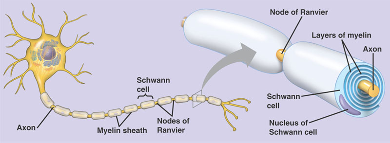
The next video is one that co-author Ken Hayworth showed me recently from his 2014 Science article on myelin sheathes surrounding single axons in the neocortex. (Besides being a senior scientist at HHMI's Janelia Farm campus, Ken is also co-founder of the Brain Preservation Foundation. (I'm a science advisor to BPF, which is trying to advance the art of brain freezing/ vitrification.)
The classic view of myelinated axons, taught for decades, is that myelin is wrapped around each axon with clockwork regularity. In between each myelinated segment is a tiny, bare node of Ranvier (free of insulation) in which the neural spike regenerates as it propagates down the long axon. This classic view is challenged in this new article. See for yourself. As the video travels down the length of an axon, myelinated areas are in green and bare areas are in brown. The unmyelinated areas are long and irregular, affording abundant opportunities for complex interactions with other neurons.
But even this challenge to classic dogma is not my main reason for showing this movie. As you travel down the axon, look to either side at the "countryside surrounding the train track." It is utterly packed with exquisite detail. This is the ultrastructure of the neuropil. This is an impenetrable forest of subcellular organelles belonging to countless, intertwined cells.
My favorite movie showing the complexity of the neuropil is this beautiful animation from Kristen Harris (U of T, Austin) and Terry Sejnowski (UCSD): Waltz through the Hippocampal Neuropil — on the synthesizer is DJ Johann S. You can also hear Terry narrate this video here, starting at minute seven of the YouTube.
Now, why is any of this important? The key question is do we really need this ultrastructural detail to replicate human cognitive phenomena in silicon? Are simple machine-learning models enough to get us human-level performance? (No!) But, this is not a question that anyone can answer now.
The answer will slowly emerge from the world's neuroscience labs and from simulation efforts by theoreticians. (Also, see my answer at the Brain Preservation Foundation. In brief, look at the most advanced AI projects that use neural nets in 2016. They're coming along but still primitive, and besides they require nuclear power plant size energy.) Meanwhile, these videos show us the incredible detail that underlies the human psyche and that may need to be replicated for realistic AI.
(Substantive comments may be emailed to bob AT bobblum DOT com. With your permission excerpts may be posted here.)
Physicist and AI theoretician Steve Omohundro responded as follows.
Bob,
Nice article! And great videos! You make an important point. It will be interesting to better understand why all that complexity is there and how much of it contributes to intelligence.
Biological systems have many requirements that computational systems mostly don't, namely
It could be that much of the complexity we see in the brain is primarily for those functions and that the portion needed for intelligence is quite simple. Or it might be that the functional behavior is intimately interwoven with the structure, and it all needs to be simulated in order to replicate intelligence.
Best, Steve


